Pellucid marginal degeneration (PMD) is a degenerative condition wherein the cornea gradually changes shape. Over time, PMD usually leads to a thinning (or ectasia) of the cornea. Patients with PMD will notice their vision deteriorate over time as the ectasia becomes more severe and widespread.Pellucid marginal degeneration is extremely similar to keratoconus, another condition that causes progressive bilateral thinning of the cornea. There two conditions are so similar they are often confused with one another.To this day, scientists are not sure whether PMD and keratoconus are different diseases or if they are two slightvariations of the same condition.No one is certain why Pellucid marginal degeneration occurs. The condition affects patients across all genders, races, and ethnicities.It sometimes manifests itself as early in the teenage years, but can also appear in the 20s, 30s, or 40s. The inconsistency and slow, gradual progression of the disease make it difficult to understand and similarly tough to diagnose.
Correcting Vision in Patients with PMD using Scleral Lenses
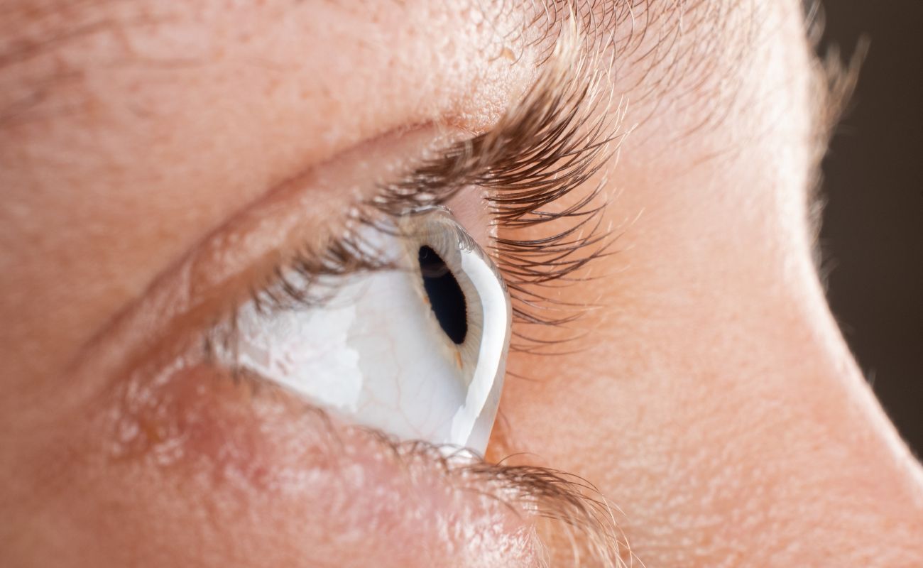
In most cases, patients with PMD use scleral lenses to correct their vision. Because Pellucid marginal degeneration results in abnormally shaped corneas, the lenses need to be custom-fitted to suit a specific patient. Standard softlenses usually do not work with PMD. Because the condition often affects 50% of the corneal surface—if not more—it can be exceedingly difficult for eye doctors to fit patients for regular contact lenses.Scleral lenses are ideal in this situation because they do not require the lens to sit on the cornea itself.Instead, the cornea is protected by a fluid-filled chamber, with the contact lens instead sitting on the sclera (the white part of the eye). As such, the high irregularity of the cornea is neutralized with the tear layer underneath the scleral lens.In addition to correcting vision in patients with PMD, scleral lenses provide the eye with the protection it needs. The fluid vault that sits on the cornea is filled with tears and saline solution, which protects the thinned and damaged tissue of the cornea. The extra protection keeps the patient comfortable while drastically improving vision. Most PMD patients wear scleral lenses during waking hours without complaining about the sensation of having something in their eye.At the Miami Contact Lens Institute, we offer scleral lens fitting for patients struggling with conditions such as PMD. If your standard contact lenses are not getting the job done, give us a call to see if scleral lenses might do the trick. You can reach our offices by calling (305) 814-2299.
Other Treatments
In most cases, scleral lenses allow Pellucid marginal degeneration patients to enjoy sharp, clear vision, maximum comfort, and overall high quality of life even as their vision slowly degenerates. Sometimes PMD can be progressiveand treatments are recommended to halt the progression. Collagen Crosslinking (CXL) has been shown to be an effective treatment for ectasia such as PMD.This minimally invasive in-office procedure is intended to strengthen the cornea by creating strong bonds (or cross-links) between corneal collagen fibers. The goal of the procedure is to prevent further progression of the PMD or ectasia. Clinical research has shown that cross-linking not only stops the progression of PMD, but it also induces flattening of the cornea and visual improvement.Collagen Crosslinking involves the instillation of riboflavin (a B vitamin) drops on the cornea after removing theepithelium. Once saturated with riboflavin, the cornea is irradiated with ultraviolet (UV) A light for about 30 minutes. The patient will likely still need to wear the specialty contact lenses or scleral lenses following the procedure.